
Nuclear Medicine Imaging/Radiology
Today, most people know that radiologists use radiation via x-rays to see inside the body and locate breaks in bones. However, less is known about how nuclear medicine works, though the two procedures are very similar.
The difference is that with nuclear medicine, the radiation is introduced into the body so that it can enter soft tissue and provide the physician with an image that they cannot obtain through traditional x-rays. Another term for nuclear imaging is radio-imaging.
How Do Radiologists Perform Radio-imaging?
The imaging department technicians inject a tiny radiotracer. The radiotracer allows the radiologist to view the surrounding tissue using the PET (Positron emission tomography) scan device. Physicians can also order that the views are superimposed with magnetic resonance imaging (MRI) or computed cosmography (CT), providing exceptional area views, also known as co-registration or image fusion.
Aside from radiologists using nuclear imaging to help diagnose diseases, procedures have been developed to allow the specialized imaging system to be used in therapy or treatments. For instance, an emerging procedure is known as radioimmunotherapy (RIT), which uses a combination of radiation therapy (brachytherapy or internal radiation) and immunotherapy to target cell activity in the immune system.
What Diseases can be Diagnosed by Radiologists using Nuclear Medicine?
Nuclear Medicine Imaging is used to diagnose
- cancer
- heart disease
- gastrointestinal disorders
- endocrine disorders
- neurological disorders
- other abnormalities in the body
Nuclear medicine has become an integral part of the diagnostic procedures for most physicians. Today, radiology departments are a large part of every diagnosis and treatment center in large part because of the benefits of nuclear medicine.
Is Nuclear Medicine Safe?
Nuclear medicine involves the use of small amounts of radioactive medications introduced into the body so that radiation technologists can gather images from within. Such images are impossible using any other methods.
The procedure involves having the patient receive the medication in advance. The medication, known as a radiopharmaceutical, may be swallowed, injected, or inhaled. Once in place, it emits low levels of radiation into nearby tissues. A gamma camera can then capture the images produced from the radioactive tissue and return essential data to the treating physician.
Naturally, many patients are concerned about ingesting anything labeled radioactive and want to know if nuclear medicine is safe. The short answer is yes. It is safe.
The long answer is that the amount of radiation in the radiotracers is minimal. In addition, because the radiation targets a specific organ of interest, the rest of the body is unaffected. Plus, radiologists take into account the patient’s body weight so that no more radiation is used than is needed for each procedure. Less radiation enters a body through nuclear medicine than a traditional x-ray, which has long been understood to be safe within carefully followed guidelines.
About Kymera Nuclear Medicine and Radiology/Imaging
The Kymera Nuclear Medicine machine is American College of Radiology (ACR) certified. ACR is at the forefront of radiology evolution, representing more than 38,000 diagnostic radiologists, radiation oncologists, interventional radiologists, nuclear medicine physicians and medical physicists.
Kymera Independent Physicians established the radiology/imaging department so that patients can obtain faster results without the inconvenience of having to leave the office.
By having our own staff of skilled radiologists, imaging techs and state-of-the-art equipment, our patients receive a better healthcare experience. Patients become better-known and the attending technicians can hold closer conferences with treating physicians. We believe this is one of the reasons we are known for providing the best cancer and other treatments in SENM.
A Kymera Radiology Technician oversees a patient procedure using PET scan equipment.

Nuclear Medicine permits targeted imaging of any part of the body.

Nuclear medicine image of the brain

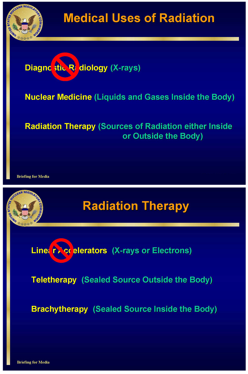
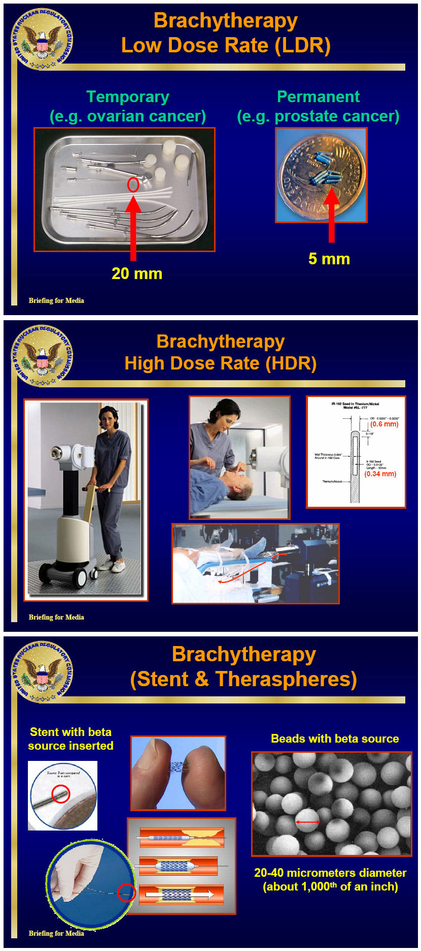
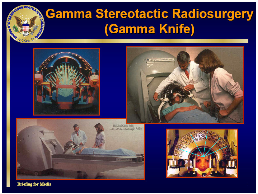
Common Nuclear Medicine Procedures
Common nuclear medicine procedures that use radioisotopes regulated by the NRC and its Agreement States include the following examples:
- Brachytherapy
- Gamma knife
- Iodine treatment for hyperthyroidism
- Portable imaging devices in dentistry and podiatry
- Bone mineral analysis
Regulatory Info from the U.S.NRC
NRC regulations are designed to ensure the proper use of radioactive materials in medical diagnosis, treatment, and research to ensure the safety of patients, medical workers, and the public, and to protect the environment.
Medical use of radioactive materials falls broadly into the two categories of diagnostic and therapeutic procedures, as described above. For additional information, see the following topics:
- The regulatory framework between the NRC and Agreement States (for contact information about regulations in your State)
- Enforcement and Incident Response
- NRC requirements in Title 10, Part 35, of the Code of Federal Regulations (10 CFR Part 35)
For Daily Health Tips and more, connect with us on Facebook @KymeraIndependentPhysicians or by clicking the image above.

Kymera…Providing A Better Healthcare Experience for over 22 Years.

